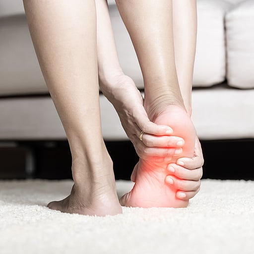
Heel pain
As you will read, heel pain is a common foot disorder that affects thousands of New Zealanders. Ask your friends - I bet one of them has suffered from, or knows someone who has suffered from this disorder.
Pain in the heel area can be caused by many factors. I hope the information within our website will enable you to better understand your condition and seek appropriate treatment. If you do have any questions please contact my rooms direct and we will do our best to help.
Signs & Symptoms
The major complaint of those with Plantar Fasciitis is pain and stiffness in the bottom of the heel. This develops gradually over time. It usually affects just one foot, but can affect both feet. Some people describe the pain as dull, while others experience a sharp pain, and some feel a burning or ache on the bottom of the foot extending outward from the heel.
The pain is usually worse in the morning when you take your first steps out of bed, or if you’ve been sitting or lying down for a while. Climbing stairs can be very difficult due to the heel stiffness. After prolonged activity, the pain can flare-up due to increased inflammation.
Pain is not usually felt during the activity, but rather just after stopping.
Causes
The Plantar Fascia, or to be more precise, the Plantar Aponeurosis, is a strong seatbelt-like ligament that runs from the bottom of the Calcaneus (heel bone) through to the bottom of the forefoot (balls of the feet). It is the most superficial band of connective tissue running along the bottom of the foot that helps support the arch.
As the arch starts to flatten and the foot absorbs the weight of the body, these structures work to maintain the arch and stabilize the foot and all help to propel us, enabling weight bearing. Normally, they accomplish this effortlessly. However, sometimes the tension stress, the load they take, is beyond their structural limit. This excessive tension can lead to damage, tearing these structures resulting in associated inflammation and pain. For this reason Plantar Fasciitis is often referred to as a “traction deformity”.
The damage/micro-injury in heel pain is often located at the proximal portion of the Plantar Aponeurosis, in particular its medial calcaneal attachment, resulting in a portion of the many micro-fibers that make up the ligamental tissue of the Plantar Aponeurosis to break/snap at this point.
If the heel pain, damage is left, the inflammation, increased blood flow, at this heel attachment can promote bone cells (osteoblasts) to start to deposit bone onto the damaged area. It is thought that this is the body’s attempt to try and strengthen the damaged area. Over time this bone cell deposition can grow and can be seen on X-ray investigations as a heel spur.
Diagnosis
Diagnosis is primarily based on history and physical examination.
Patients may present with heel pain with their first steps in the morning or after prolonged sitting, and sharp pain with palpation of the medial plantar calcaneal region. Discomfort in the proximal plantar fascia can be elicited by passive ankle/first toe dorsi flexion.
Diagnostic imaging is rarely needed for the initial diagnosis of Plantar Fasciitis. Use of ultrasonography and magnetic resonance imaging (MRI) is reserved for recalcitrant cases or to rule out other heel pathology.
Ultrasonography findings that support the diagnosis of Plantar Fasciitis include proximal plantar fascia thickness greater than 4 mm and areas of hypo-echogenicity.
Magnetic Resonance Imaging (MRI), although expensive, can be a valuable tool for assessing causes of recalcitrant heel pain. Diagnostic findings include increased proximal plantar fascia thickening with increased signal intensity on t2-weighted and short tau inversion recovery images.
Treatment
Our podiatrist is able to provide a range of effective, non-invasive treatments aimed at providing pain relief and resolving the cause of your heel pain.
These can include:
Recommendation to reduce activities to enable the tissues within the heel to repair.
Sometimes the use of an ice pack can also offer pain relief.
The use of non-steroidal anti-inflammatory medications can also offer relief, however it is important that you consult your GP or contact us before you commence using this type of medication.
Specifically, targeting stretching programmes.
Specialists' taping techniques.
The use of night splints is aimed at stretching abnormally tight calf muscles that can cause heel pain.
Orthotic treatment offers pain relief by reducing abnormal stresses within the foot.
Extra-corporeal Shockwave Therapy (ESWT) – Focal shockwave as opposed to the more commonly used “jack-hammer” low energy radial pulse wave therapy.
Ultrasonically guided glucocorticoid steroid injection utilising local analgesia.
In severe cases, the use of a non-weight bearing air cast boot may be used to enabled the inflamed area to heal, normally returning to walking anywhere from 2-6 weeks.
Patients can often present to our clinic after trying to use off-the-shelf shoe insert devices, eg gel heel cups, or soft “shock absorbing” inserts and footwear. Under the load of body weight, these soft type devices and footwear, create an unstable platform/foundation causing the foot to wobble, move too much under body weight and in doing so, increase the abnormal stresses within the foot, failing to offer pain relief.
The successful treatment of heel pain depends upon a sound diagnosis.
Prevention
Stretching is the best non-invasive treatment for Plantar Fasciitis. Gently stretching your calf muscles will help to reduce the tension on your Achilles Tendons and plantar fascia. These are the main stay of self-treatment and help to reduce the risk of developing heel pain.
In one exercise, you lean forward against a wall with one knee straight and heel on the ground. Your other knee is bent. Your heel cord and foot arch stretch as you lean. Hold for 10 seconds, relax and straighten up. Repeat 20 times for each sore heel. It is important to keep the knee fully extended on the side being stretched.
Perform at least three sets of stretches per day. You cannot perform the stretch too often. The most important times to stretch are before taking the first step in the morning and before standing after a period of prolonged sitting.