Ankle Injuries
The ankle joint is comprised of three bones: The Talus that is located within the rear foot, sitting on top of the Talus, the Fibula and the Tibia (from the leg). The three bones together form a mortise (on the top of the talus), as well as two joint areas (on the inside and outside of the ankle), the Tibial and Fibular syndesmosis, sometimes called the "gutters."
The ankle allows the foot to move upwards and downwards, often referred to as ankle flexion and extension.
The foot also needs to roll in (pronation) and roll outwards (supination). Pronation and supination is enabled by the joint that lays beneath the ankle joint, it’s called the subtalar joint. The subtalar joint is a very important joint as it allows us to stand upright and walk on two legs. It provides shock absorption during pronation and acts like a torque converter, allowing the leg, thigh and pelvis to rotate inwards and outwards during your gait cycle. Rotating outwards during supination pushes together the joints in the foot “locking the foot” creating a rigid lever effect in the foot, allowing the foot to push efficiently during the propulsive stage of gait.
These joints are surrounded by a joint capsule, referred to as a synovial membrane or synovium. The synovial membrane produces synovial fluid, a clear slippery joint fluid that lubricates the joint. The synovial membrane also contains blood vessels that provide oxygen and nutrients to the joint. The ankle and subtalar joint also include ligaments that hold them together. Ligaments are non-elastic fibrous connective tissues that cross joint spaces and anchor into the adjacent bone providing intrinsic joint stability. They prevent joints from moving beyond their normal range, beyond their cartilaginous, articular surfaces. Ligaments are normally incredibly strong.
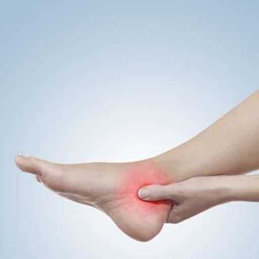
Signs & Symptoms
Ankle injuries can be debilitating, seriously affecting your quality of life, and literally stopping you in your tracks.
Injuries can cause pain and sometimes, but not always, swelling in the ankle joint or subtalar joint. Pain on movement can be continual or intermittent. Patients can complain of severe, sharp type pain that “catches you”. It can seem to appear from nowhere while walking. Sometimes the ankle or subtalar joint can jam / lock or suddenly roll outwards causing you to stumble or fall. Moving the ankle upwards, extending it when crouching down can also aggravate the ankle injury causing a sharp type pain. Walking on uneven or across angled, sloping type surface can also produce pain within an injured ankle or subtalar joint.
Causes
An ankle injury can occur when one rolls over a rock, lands off a curb, or steps in a small hole or crack in the road. Usually, the sprain is only mild, but on occasion, it may seriously injure the joint capsule, synovial membrane and ligaments or tendons surrounding the ankle joint.
Most ankle sprains involving the ligaments are weight-bearing injuries. When a foot rolls outward (supinates) and the front of the foot points downwards as he or she lands on the ground, a lateral ankle sprain can be a result.
It is usually this situation that causes injury to the anterior talofibular ligament. However, when the foot rolls inwards (pronates) and the forefoot turns outward (abducts), the ankle is subject to an injury involving the deltoid ligament that supports the outside of the ankle.
Other injuries to the ankle and subtalar joint can be caused by wear and tear, causing damage to the articular surfaces of the joints, wearing away the cartilage resulting in inflammation and pain. This is called osteoarthritis.
Sometimes the foot rolls in too much (excessive pronation) causing the outside borders of the subtalar joint to pinch or impinge. This can cause a reactive bony ridge to form, producing further inflammation and pain. This is called a subtalar coalition. This can also occur at the articular borders of the ankle joint.
Other causes of ankle and subtalar joint pain can include but is not limited to:
Inflammatory conditions including: Rheumatoid, Systemic Lupus Erythematosus, Gout, Pseudo Gout, Psoriatic arthritis, scleroderma, Charcot Marie Tooth disorder.
Infection can also cause ankle and subtalar joint pain. This can include, cellulitis, osteomyelitis, necrotising fasciitis, septic arthritis
Diagnosis
As with all foot and ankle conditions, an early and accurate diagnosis is the key to a successful treatment.
When assessing an ankle injury we will want to know the mechanism of injury and history of previous ankle sprains. Where the foot was located at the time of injury, "popping" sensations, and whether you can put weight on the ankle are all important questions that need to be asked.
The physical examination can help confirm a suspected diagnosis, based on the history of the injury. A careful, hands on approach can be an effective way to determine exactly where the injury is located e.g. the ankle or subtalar joint, the front, side or rear of the joint. A thorough visual assessment looks for any obvious deformities of the ankle or foot, black and blue discolouration, swelling, or disruption of the skin.
Ankle instability is evaluated while weight-bearing. Manual muscle testing is also valuable when checking for ankle instability. One of the more critical tests that you should be able to perform before allowing the resumption of activity is a "single toe raise" test. If you are unable to do this, we might suspect ligamentous injury or ankle instability.
When crackling, extreme swelling and tenderness are present, together with a limited range of motion, we may suspect a fracture of the ankle.
X-rays help but do not rule out fractures, degenerative joint disease, osteochondral defect (OCD) or osteochondral lesion of the talus (OLT). “Osteo” means bone and “chondral” refers to cartilage. The use of a MRI or Bone Scan (Skeletal Scintigraphy) can be useful to help diagnose these types of lesions. Stress X-rays and fluoroscopy (a continual X-ray that can record movement) are taken when ligamentous rupture or ankle instability are suspected.
A diagnostic local anaesthetic block, isolating the ankle joint or the affected area is useful to diagnose the injury, it is also useful to rule out damage or injury to the Subtalar joint that is positioned just below the ankle joint and can be overlooked when diagnosing this type of injury.
Treatment
Management of this injury relies on early and accurate diagnosis. The treatment of an ankle injury depends of the location and cause and severity of the injury.
Common first aid treatment can include:
Rest, Ice pack in a towel, Compression and Elevation, above your heart level (R.I.C.E.)
If the injury is limited to slight stretching of a ligament and synovial membrane, the short term, 4 to 6 week use of sports strapping techniques, e.g. the stirrup strapping technique may help.
The 4 to 6 week use of compressional non-adhesive bandaging and compression or lace up ankle bracing can be helpful.
The short-term use of non-steroidal anti-inflammatories (NSAIDs) can also provide short-term pain relief . Please ensure you receive professional advice from your Podiatrist or GP before your commence the use of anti-inflammatory medication.
If a more severe injury is diagnosed, a partial ligament tear, a 6 to 8 week use of a Controlled Ankle Motion (CAM) Boot may also be recommended to help you walk while you recover.
If a fracture is diagnosed the use of a non- weight bearing Fracture Boots or Aircast boot maybe used. This also depends of the type of fracture and in some cases, hospitalisation and surgical treatment may be required.
Long-term treatment is aimed at rehabilitation of your ankle joint, returning it to a normal pain-free function. This can include the use of a regime of strengthening exercise targeted at increasing the power of the muscles in your leg that control the stability of your foot and ankle.
Exercises that help your brain improve its function to undertake proprioception are useful. They help perceive “feel it” when your foot and ankle are unstable, enabling you to use the muscles that stabilise your ankle.
The use of prescription orthotics can also prove useful. They can help support your foot, effectively making it harder for you to move your foot in the direction of an ankle, reducing the tension from the injured tissues. This can reduce excessive inflammation at the site of injury, provide pain relief and in doing so aid recovery.
Extra corporeal shockwave therapy (ESWT) is an intervention that stimulates the body’s natural healing process. Additionally, SWT has been shown to have a direct effect on local nerve endings resulting in a decrease in pain.
Shockwaves stimulate fibroblasts which are cells responsible for the healing of connective tissue such as tendons.
Diminishes pain by two mechanisms:
Hyper-stimulation anaesthesia - local nerve endings are overwhelmed with so many stimuli that their activity diminishes, resulting in a short-term reduction in pain
Gate-control mechanism - whereby local nerves are stimulated to recalibrate perception of pain and result in a longer-term reduction in pain.
Sometimes a muscular imbalance in your legs can make your ankle inherently unstable and the daily use of a targeting stretching regime or stretching splint may be recommended.
Prevention
Please remember, when an acute or chronic ankle sprain is not treated, as unfortunately is all too often the case, repeated ankle sprains may occur. Because chronic ankle injuries do not show acute inflammation even when the ankle is weak and unstable, this may set you up for another ankle sprain when least suspected. A successive sprain may be more severe than the first, and cause an even more significant injury.
The most important point to keep in mind when talking about ankle injuries, then, is to prevent the condition from becoming chronic or recurrent.
Exercises to help increase the strength of the muscles in your leg that stabilise your ankle are helpful e.g. learning to balance on a wobble board.
Sometimes the shape of your foot can cause it to move in the direction of an ankle sprain. The use of prescription orthotics can support your foot, making it difficult for your foot to move in the direction of an ankle sprain, commonly supinating to an “inverted” position.
If you have pain or swollen ankles during activities e.g. standing, walking, hiking, cycling, skiing or running, please don't ignore early warning signs. If you have any questions, please contact us and we will do our best to help.